Who Let the Microbes Out: A Paw Print of Doggy Skin Bacteria
A house is not a home without a dog, and a dog isn’t a “D-O-double-G” without its microbial “crew.”
Human microbiome research is progressing rapidly, and we are always learning how the bacteria living on and inside of us contribute to our survival and well-being. Although we are making some headway to understanding the role of human flora in our bodies and in disease progression, we know far less about the microbial flora in our pooch friends.
The diverse microbial landscape on skin is essential for all animals, as it helps maintain essential oils and plays “guard” in the first line of immune defense. However, living on animal skin isn’t easy—it’s exposed to the world and all its creatures, and endures some serious physical contact with them. There are many skin diseases that may alter skin microbe colonization, such as atopic dermatitis (AD), one of the most common skin infections in dogs (and people). Dogs that do develop AD have an increased sensitivity to many allergens. Additionally, many dogs with allergic AD are subject to bacterial skin infections. With this in mind, a group of researchers at Texas A&M recently published a study in PLOS ONE where they identify the microbial makeup of dog skin. The authors “hired” a crew of healthy dogs, as well as a group of dogs with allergic AD, to characterize the differences in their bacterial communities.
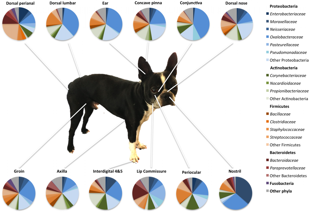
To obtain samples of as many skin-inhabiting critters as possible, the researchers brushed swabs back and forth on 12 different skin sites from the “healthy” dog squad, and 4 different skin sites from the allergic AD dogs, with the sites shown in the image below.
After extracting and sequencing the DNA from the swab culture samples, the authors were able to characterize the microbial inhabitants of the dogs’ skin for each site, and compare the species distribution found on the healthy dogs’ skin to those found on the allergic dogs. In healthy dogs, the lowest abundance of species, also called species richness, were found in their mucosal areas, such as the nostrils; whereas the haired-skin areas, such as the groin and axilla, had the highest species richness. Notably, ears took the “bone” with 866 different species present!
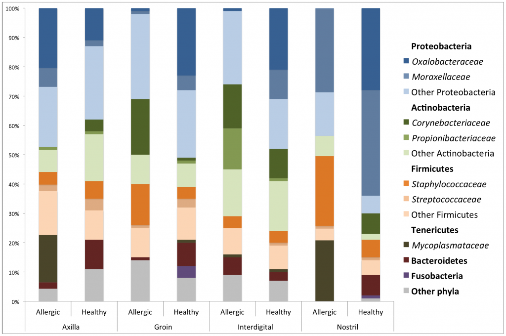
Among all dogs tested, the authors found a total of 17 different phyla, with Proteobacteria being the most abundant phylum. Ralstonia was the most abundant genus, likely because it’s found in dirt and water, the favorited doggy playground. Although there was high variability among the individual dogs tested, the authors found that the proportion of Ralstonia was much lower in all allergic dogs.
The species richness was much lower in the allergic dogs as compared to healthy dogs, as seen in the image above. The authors suggest that this difference may be the result of the antimicrobial washes that allergic dogs are often prescribed by vets for treatment, as well as skin changes induced by the allergic inflammation response.
While there is much work to be done characterizing the full doggie microbiome, and this study had a limited sample size, the authors have made steps toward characterizing the bacterial ecology of dog skin, and discovered that dog skin may contain much richer and more diverse microbial communities than were previously suggested. Like with the human microbiome, researchers might now investigate the role of the dog microbiome in normal function, disease progression, and better treatments for our favorite co-pilots. So next time you let the dogs out, give a thought to their bacterial companions!
Citation: Rodrigues Hoffmann A, Patterson AP, Diesel A, Lawhon SD, Ly HJ, et al. (2014) The Skin Microbiome in Healthy and Allergic Dogs. PLoS ONE 9(1): e83197. doi:10.1371/journal.pone.0083197
Image 1: Hawaiian Burton by Sarah Nichols
Images 2-3: Figures 4 and 7 from the published article.


